Newly Discovered Neural Network Gets Visual and Motor Circuits in Sync
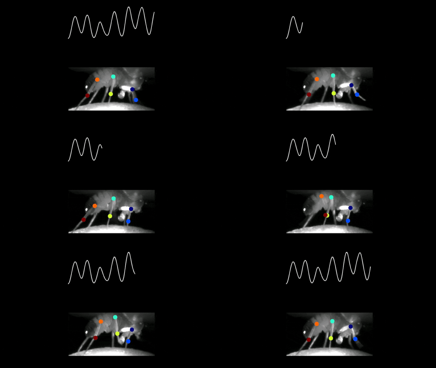
A fruit fly strolls on a little styrofoam ball made into a drifting 3D treadmill. The space is totally dark, and yet, an electrode recording visual nerve cells in the fly’s brain passes on a mystical stream of neural activity, fluctuating like a sinusoidal wave.
When Eugenia Chiappe, a neuroscientist at the Champalimaud Foundation in Portugal, very first saw these outcomes, she had an inkling her group had actually made a remarkable discovery. They were tape-recording from visual nerve cells, however the space was dark, so there was no visual signal that might drive the nerve cells because way.
“That meant that the unusual activity was either an artifact, which was unlikely, or that it was coming from a non-visual source,” Chiappe remembered. “After the possibility of interference was investigated and dismissed, I was sure: the neurons were faithfully tracking the animal’s steps.”
A couple of years and lots of brand-new insights later on, Chiappe and her group now provide their discovery in the clinical journal Neuron: a bi-directional neural network linking the legs and the visual system to form walking.
“One of the most amazing elements of our finding is t hat this network supports strolling on 2 various timescales at the same time,” statedChiappe “It operates on a fast timescale to monitor and correct each step while promoting the animal’s behavioral goal.”
The charge of a visual nerve cell (top) in the brain of a fruit fly was tape-recorded while the animal was strolling easily on top of a drifting 3D treadmill. Tracking the position of the legs reveals that the charge is in tune with the fly’s front leg. Credit: Terufumi Fujiwara & & Eugenia Chiappe, Champalimaud Foundation
Tracking neural “mood”
“Vision and action may seem unrelated, but they are actually tightly associated; just choose a point on the wall and try placing your finger on it with your eyes closed,” statedChiappe “Still, little is known about the neural basis of this link.”
In this research study, the group concentrated on a specific kind of visual nerve cell that is understood to link to motor brain locations. “We wanted to identify the signals that these neurons receive and understand if and how they participate in movement,” described Terufumi Fujiwara, the very first author of the research study.
To response these concerns, Fujiwara utilized an effective strategy called whole-cell spot recording that allowed him to take advantage of the nerve cells’ “mood,” which can be either favorable or unfavorable.
“Neurons communicate with each other using electric currents that alter the overall charge of the receiving neuron. When the neuron’s net charge is more positive, it is more likely to become active and then transmit signals to other neurons. On the other hand, if the charge is more negative, the neuron is more inhibited,” Fujiwara described.
Watching each action
The group tracked the nerve cells’ charge and exposed that it was synched to the animal’s actions in a way that was optimum for fine-tuning each motion.
“When the foot was up in the air, the neuron was more positive, ready to send out adjustment directions to the motor region if needed. On the other hand, when the foot was on the ground, making adjustments impossible, the charge was more negative, effectively inhibiting the neuron,” stated Chiappe.
Keeping the course
When the group evaluated their outcomes even more, they observed that charge of the nerve cells was likewise altering on a longer timescale. Specifically, when the fly was strolling quick, the charge ended up being progressively a growing number of favorable.
“We believe that this variation helps maintain the animal’s behavioral goal,” statedFujiwara “The longer the fly has been walking fast, the higher are the chances that it would need help to maintain this action plan. Therefore, the neurons become increasingly ‘more alert’ and ready to be recruited for movement control.”
The brain is not constantly the one in charge
Many experiments followed, developing a fuller description of the network and showing its direct participation in strolling. But according to Chiappe, this research study goes even further than exposing a brand-new visual-motor circuit, it likewise supplies a fresh point of view on the neural systems of motion.
“The current view of how behavior is generated is very ‘top-down’: the brain commanding the body. But our results provide a clear example of how signals originating from the body contribute to movement control. Though our findings were made in the fly animal model, we speculate that similar mechanisms may exist in other organisms. Speed-related representations are critical during exploration, navigation, and spatial perception, functions that are common to many animals, including humans,” she concluded.
Reference: “Walking strides direct rapid and flexible recruitment of visual circuits for course control in Drosophila” 6 May 2022, Neuron
DOI: 10.1016/ j.neuron.202204008


