MIT scientists have actually discovered areas of the baby visual cortex that reveal strong choices for either deals with, bodies, or scenes, simply as they perform in grownups. Credit: MIT News, with images thanks to the scientists and iStockphoto
Study recommends this location of the visual cortex emerges much previously in advancement than formerly believed.
Within the visual cortex of the adult brain, a little area is specialized to react to faces, while neighboring areas reveal strong choices for bodies or for scenes such as landscapes.
Neuroscientists have actually long assumed that it takes several years of visual experience for these locations to establish in kids. However, a brand-new MIT research study recommends that these areas form much earlier than formerly believed. In a research study of children varying in age from 2 to 9 months, the scientists recognized locations of the baby visual cortex that currently reveal strong choices for either deals with, bodies, or scenes, simply as they perform in grownups.
“These data push our picture of development, making babies’ brains look more similar to adults, in more ways, and earlier than we thought,” states Rebecca Saxe, the John W. Jarve Professor of Brain and Cognitive Sciences, a member of MIT’s McGovern Institute for Brain Research, and the senior author of the brand-new research study.
Using practical magnetic resonance imaging (fMRI), the scientists gathered functional information from more than 50 babies, a far higher number than any research study laboratory has actually had the ability to scan previously. This enabled them to analyze the baby visual cortex in such a way that had actually not been possible previously.
“This is a result that’s going to make a lot of people have to really grapple with their understanding of the infant brain, the starting point of development, and development itself,” states Heather Kosakowski, an MIT college student and the lead author of the research study, which was released on November 15, 2021, in Current Biology
Distinctive areas
More than 20 years earlier, Nancy Kanwisher, the Walter A. Rosenblith Professor of Cognitive Neuroscience at MIT, utilized fMRI to find the fusiform face location: a little area of the visual cortex that reacts far more highly to faces than any other type of visual input.
Since then, Kanwisher and her associates have actually likewise recognized parts of the visual cortex that react to bodies (the extrastriate body location, or EBA), and scenes (the parahippocampal location location, or PPA).
“There is this set of functionally very distinctive regions that are present in more or less the same place in pretty much every adult,” states Kanwisher, who is likewise a member of MIT’s Center for Brains, Minds, and Machines, and an author of the brand-new research study. “That raises all these questions about how these regions develop. How do they get there, and how do you build a brain that has such similar structure in each person?”

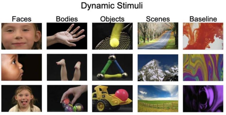
After entering into the specialized scanner, in addition to a moms and dad, the children enjoyed videos that revealed either deals with, body parts such as kicking feet or waving hands, things such as toys, or natural scenes such as mountains. This figure reveals examples of the images utilized in the research study. Credit: Courtesy of the scientists
One method to attempt to respond to those concerns is to examine when these extremely selective areas very first establish in the brain. A longstanding hypothesis is that it takes a number of years of visual experience for these areas to slowly end up being selective for their particular targets. Scientists who study the visual cortex have actually discovered comparable selectivity patterns in kids as young as 4 or 5 years of ages, however there have actually been couple of research studies of kids more youthful than that.
In 2017, Saxe and among her college student, Ben Deen, reported the very first effective usage of fMRI to study the brains of awake babies. That research study, that included information from 9 children, recommended that while babies did have locations that react to faces and scenes, those areas were not yet extremely selective. For example, the fusiform face location did disappoint a strong choice for human faces over every other type of input, consisting of bodies or the faces of other animals.
However, that research study was restricted by the little number of topics, and likewise by its dependence on an fMRI coil that the scientists had actually established specifically for children, which did not provide as high-resolution imaging as the coils utilized for grownups.

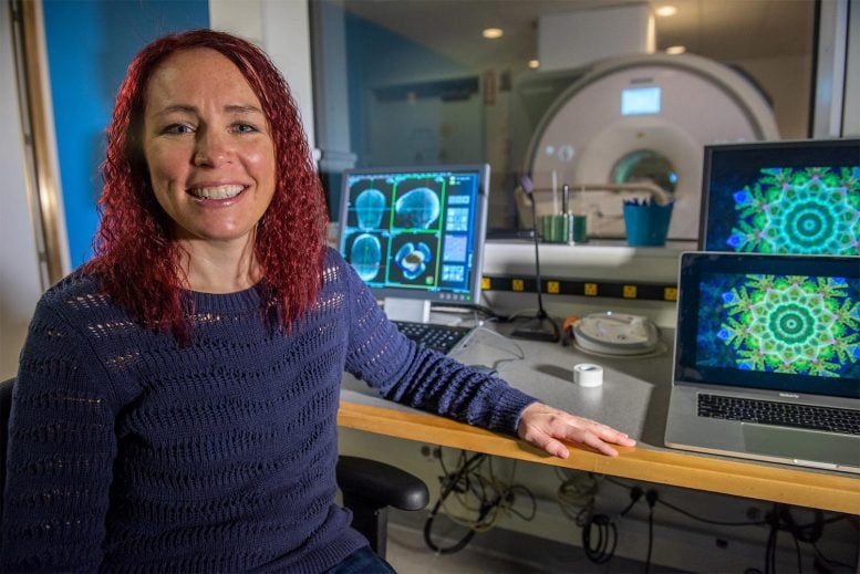
“This is a result that’s going to make a lot of people have to really grapple with their understanding of the infant brain, the starting point of development, and development itself,” states Heather Kosakowski, visualized, an MIT college student and the lead author of the research study. Credit: Kris Brewer
For the brand-new research study, the scientists wished to attempt to improve information, from more children. They constructed a brand-new scanner that is more comfy for children and likewise more effective, with resolution comparable to that of fMRI scanners utilized to study the adult brain.
After entering into the specialized scanner, in addition to a moms and dad, the children enjoyed videos that revealed either deals with, body parts such as kicking feet or waving hands, things such as toys, or natural scenes such as mountains.
The scientists hired almost 90 children for the research study, gathered functional fMRI information from 52, half of which contributed higher-resolution information gathered utilizing the brand-new coil. Their analysis exposed that particular areas of the baby visual cortex program extremely selective reactions to faces, body parts, and natural scenes, in the very same areas where those reactions are seen in the adult brain. The selectivity for natural scenes, nevertheless, was not as strong when it comes to faces or body parts.
The baby brain
The findings recommend that researchers’ conception of how the baby brain establishes might require to be modified to accommodate the observation that these specialized areas begin to look like those of grownups earlier than anybody had actually anticipated.
“The thing that is so exciting about these data is that they revolutionize the way we understand the infant brain,” Kosakowski states. “A lot of theories have grown up in the field of visual neuroscience to accommodate the view that you need years of development for these specialized regions to emerge. And what we’re saying is actually, no, you only really need a couple of months.”
Because their information on the location of the brain that reacts to scenes was not as strong when it comes to the other areas they took a look at, the scientists now prepare to pursue extra research studies of that area, this time revealing children images on a much bigger screen that will more carefully simulate the experience of being within a scene. For that research study, they prepare to utilize near-infrared spectroscopy (NIRS), a non-invasive imaging method that does not need the individual to be inside a scanner.
“That will let us ask whether young babies have robust responses to visual scenes that we underestimated in this study because of the visual constraints of the experimental setup in the scanner,” Saxe states.
The scientists are now more evaluating the information they collected for this research study in hopes of discovering more about how advancement of the fusiform face location advances from the youngest children they studied to the earliest. They likewise intend to carry out brand-new experiments taking a look at other elements of cognition, consisting of how children’ brains react to language and music.
Reference: “Selective responses to faces, scenes, and bodies in the ventral visual pathway of infants” by Heather L. Kosakowski, Michael A. Cohen, Atsushi Takahashi, Boris Keil, Nancy Kanwisher and Rebecca Saxe, 15 November 2021, Current Biology
DOI: 10.1016/ j.cub.202110064
The research study was moneyed by the National Science Foundation, the National Institutes of Health, the McGovern Institute, and the Center for Brains, Minds, and Machines.


