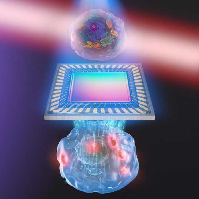
An creative representation of the brand-new imaging technique called biochemical quantitative stage imaging with midinfrared photothermal result, established by a research study group at the University of Tokyo. Credit: s-graphics.co.jp
No damage brought on by strong light, no synthetic dyes or fluorescent tags required.
The within living cells can be seen in their natural state in higher information than ever prior to utilizing a brand-new method established by scientists in Japan. This advance must assist expose the complex and delicate biological interactions of medical secrets, like how stem cells establish or how to provide drugs better.
“Our system is based on a simple concept, which is one of its advantages,” stated Associate Professor Takuro Ideguchi from the University of Tokyo Research Institute for Photon Science and Technology. The outcomes of Ideguchi’s group were released just recently in Optica, the Optical Society’s research study journal.
The brand-new technique likewise has the benefits of not requiring to eliminate the cells, harm them with extreme light, or synthetically connect fluorescent tags to particular particles.
The method integrates 2 pre-existing microscopy tools and utilizes them at the same time. The mix of these tools can be thought about just as like a coloring book.
“We gather the black-and-white outline of the cell and we virtually color in the details about where different types of molecules are located,” stated Ideguchi.
Quantitative stage microscopy collects info about the black-and-white summary of the cell utilizing pulses of light and determining the shift in the light waves after they travel through a sample. This info is utilized to rebuild a 3D picture of the significant structures inside the cell.
Molecular vibrational imaging offers the virtual color utilizing pulses of mid-infrared light to include energy to particular kinds of particles. That additional energy triggers the particles to vibrate, which warms up their regional environments. Researchers can select to raise the temperature level of particular kinds of chemical bonds by utilizing various wavelengths of midinfrared light.
Researchers take a quantitative stage microscopy picture of the cell with the midinfrared light shut off and an image with it switched on. The distinction in between those 2 images then exposes both the summary of significant structures inside the cell and the specific areas of the kind of particle that was targeted by the infrared light.
Researchers describe their brand-new combined imaging technique as biochemical quantitative stage imaging with mid-infrared photothermal result.
“We were impressed when we first observed the molecular vibrational signature characteristic of proteins, and we were further excited when this protein-specific signal appeared in the same location as the nucleolus, an intracellular structure where high amounts of proteins would be expected,” stated Ideguchi.
Ideguchi’s group hopes their method may permit scientists to figure out the circulation of basic kinds of particles inside single cells. The quantitative stage microscopy summary of significant structures might be practically colored in utilizing various wavelengths of light to particularly target proteins, lipids (fats) or nucleic acids (DNA, RNA).
Currently, catching one total image can take 50 seconds or longer. Researchers are positive that they can accelerate the procedure with easy enhancements to their tools, consisting of a higher-powered source of light and a more delicate video camera.
Reference: “Label-free biochemical quantitative phase imaging with mid-infrared photothermal effect” by Miu Tamamitsu, Keiichiro Toda, Hiroyuki Shimada, Takaaki Honda, Masaharu Takarada, Kohki Okabe, Yu Nagashima, Ryoichi Horisaki and Takuro Ideguchi, 20 April 2020, Optica.
DOI: 10.1364/OPTICA.390186
Collaborators at Osaka University, other departments at the University of Tokyo and the Japan Science and Technology Agency likewise added to this research study.


