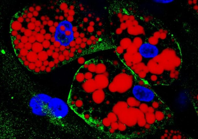
Cultured separated adipocytes: lipids are stained red; the protein ACE-2, which acts as the receptor for SARS-CoV-2, is green; cell nuclei are blue. Credit: Amanda Passos & & Fl ávio Protasio Veras
Two kinds of adipocytes (fat cells) were contaminated in the lab: one acquired from human stem cells separated from subcutaneous tissue and the other separated from stem cells drawn from visceral fat.
Experiments reveal that visceral fat– fat around the liver, intestinal tracts, and other organs, thought about a danger element for heart disease, diabetes, and hypertension– contributes more to extreme


