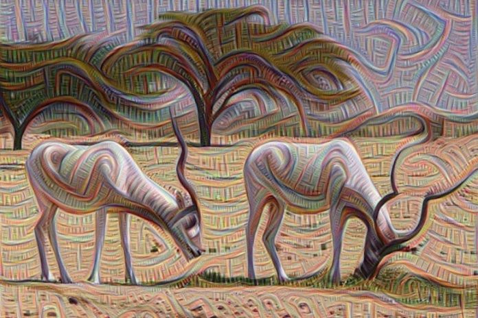
An ibis as “seen” by a device, 2015. This processed image, which is based upon a photo by Dr. Zachi Evenor, is thanks to software application engineer Guenther Noack, 2015. Credit: Dr. Guenther Noack, 2015, replicated from Wikimedia Commons (CC BY 4.0).
Medical University of South Carolina scientists report in Current Biology that the brain utilizes comparable visual locations for psychological images and vision, however it utilizes low-level visual locations less specifically with psychological images than with vision. These findings include understanding to the field by refining approaches to study psychological images and vision. In the long-lasting, it might have applications for psychological health conditions impacting psychological images, such as trauma. One sign of PTSD is invasive visual pointers of a terrible occasion. If the neural function behind these invasive ideas can be much better comprehended, much better treatments for PTSD might possibly be established.
The research study was carried out by an MUSC research study group led by Thomas P. Naselaris, Ph.D., associate teacher in the Department of Neuroscience. The findings by the Naselaris group aid respond to an olden concern about the relationship in between psychological images and vision.
“We know mental imagery is in some ways very similar to vision, but it can’t be exactly identical,” discussed Naselaris. “We wanted to know specifically in which ways it was different.”
“When you’re imagining, brain activity is less precise. It’s less tuned to the details, which means that the kind of fuzziness and blurriness that you experience in your mental imagery has some basis in brain activity.” — Dr. Thomas Naselaris
“There’s this brain-like artificial system, a neural network, that synthesizes images,” Naselaris discussed. “It’s like a biological network that synthesizes images.”
The Naselaris group trained this network to see images and after that took the next action of having the computer system envision images. Each part of the network resembles a group of nerve cells in the brain. Each level of the network or nerve cell has a various function in vision and after that psychological images.
To test the concept that these networks resemble the function of the brain, the scientists carried out an MRI research study to see which brain locations are triggered with psychological images or vision.
While inside the MRI, individuals saw images on a screen and were likewise asked to envision images at various points on the screen. MRI imaging made it possible for scientists to specify which parts of the brain were active or peaceful while individuals saw a mix of animate and inanimate things.
Once these brain locations were mapped, the scientists compared the arise from the computer system design to human brain function.

The picture of an ibis by Dr. Zachi Evenor on which the computer-processed image is based. Credit: Dr. Zachi Evenor, Reproduced from Wikimedia Commons (CC BY 4)
They found that both the computer system and human brains worked likewise. Areas of the brain from the retina of the eye to the main visual cortex and beyond are both triggered with vision and psychological images. However, in psychological images, the activation of the brain from the eye to the visual cortex is less exact, and in a sense, diffuse. This resembles the neural network. With computer system vision, low-level locations that represent the retina and visual cortex have exact activation. With psychological images, this exact activation ended up being diffuse. In brain locations beyond the visual cortex, the activation of the brain or the neural network is comparable for both vision and psychological images. The distinction depends on what’s occurring in the brain from the retina to the visual cortex.
“There’s this brain-like artificial system, a neural network, that synthesizes images. It’s like a biological network that synthesizes images.” — Dr. Thomas Naselaris
“When you’re imagining, brain activity is less precise,” stated Naselaris. “It’s less tuned to the details, which means that the kind of fuzziness and blurriness that you experience in your mental imagery has some basis in brain activity.”
Naselaris hopes these findings and advancements in computational neuroscience will cause a much better understanding of psychological health problems.
The fuzzy dream-like state of images assists us to compare our waking and dreaming minutes. In individuals with PTSD, intrusive pictures of distressing occasions can end up being devastating and seem like truth in the minute. By understanding how psychological images works, researchers might much better comprehend mental disorders identified by interruptions in psychological images.
“When people have really invasive images of traumatic events, such as with PTSD, one way to think of it is mental imagery dysregulation,” discussed Naselaris. “There’s some system in your brain that keeps you from generating really vivid images of traumatic things.”
“The extent to which the brain differs from what the machine is doing gives you some important clues about how brains and machines differ. Ideally, they can point in a direction that could help make machine learning more brainlike.” — Dr. Thomas Naselaris
A much better understanding of how this operates in PTSD might offer insight into other psychological health issue identified by psychological images interruptions, such as schizophrenia.
“That’s very long term,” Naselaris clarified.
For now, Naselaris is concentrating on how psychological images works, and more research study requires to be done to attend to the connection to psychological health.
A restriction of the research study is the capability to recreate completely the psychological images conjured by individuals throughout the experiment. The advancement of approaches for equating brain activity into viewable images of psychological images is continuous.
This research study not just checked out the neurological basis of seen and pictured images however likewise set the phase for research study into enhancing expert system.
“The extent to which the brain differs from what the machine is doing gives you some important clues about how brains and machines differ,” stated Naselaris. “Ideally, they can point in a direction that could help make machine learning more brainlike.”
###
Reference: “Generative Feedback Explains Distinct Brain Activity Codes for Seen and Mental Images” by Jesse L. Breedlove, Ghislain St-Yves, Cheryl A. Olman and Thomas Naselaris, 30 April 2020, Current Biology.
DOI: 10.1016/j.cub.2020.04.014
Catherine Bridges is an M.D., Ph.D. trainee at MUSC who operates in the lab of Dr. Christopher Cowan.


