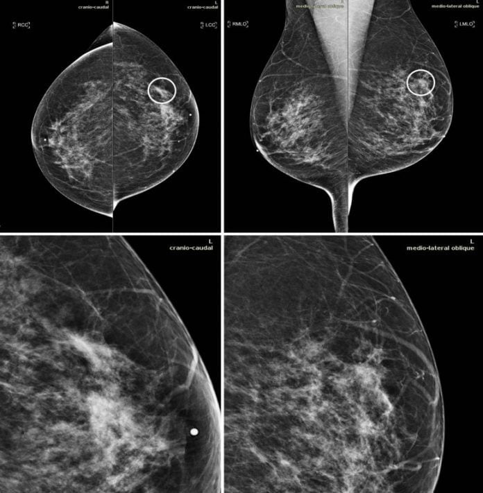
Mammograms in a 51-year-old lady with intrusive ductal cancer. The upper panels reveal the craniocaudal and the mediolateral oblique views. The lower panels reveal a close-up of the left breast location including the sore. The case is among the false-negative cases consisted of in the dataset. Accordingly, the preliminary screening evaluation was a BI-RADS 2, implying noticeable findings were evaluated as benign. After 1 year, the client provided for another screening evaluation. This time, a focal asymmetry with associated distortion within the left breast was discovered; the client was remembered and detected with a 1.5-cm mass in the upper external quadrant of the left breast on the craniocaudal view (circle). Credit: Radiological Society of North America
Artificial intelligence (AI) can boost the efficiency of radiologists in checking out breast cancer screening mammograms, according to a research study released in Radiology: Artificial Intelligence.
Breast cancer screening with mammography has actually been revealed to enhance diagnosis and decrease death by identifying illness at an earlier, more treatable phase. However, numerous cancers are missed on screening mammography, and suspicious findings typically end up being benign. An earlier research study from Radiology discovered that, typically, just 10% of females remembered from evaluating for extra diagnostic workup based upon suspicious findings are eventually discovered to have cancer.
AI-based algorithms represent an appealing opportunity for enhancing the precision of digital mammography. Developers “train” the AI on existing images, teaching it to acknowledge irregularities related to cancer and identify them from benign findings. The programs can then be evaluated on various sets of images. AI uses not just the possibility of much better cancer detection however likewise enhanced performance for radiologists.
For the research study, scientists utilized MammoScreen, an AI tool that can be used with mammography to help in cancer detection. The AI system is developed to determine areas suspicious for breast cancer on 2D digital mammograms and evaluate their probability of malignancy. The system takes as input the total set of 4 views making up a mammogram and outputs a set of image positions with an associated suspicion rating.
Fourteen radiologists evaluated a dataset of 240 2D digital mammography images obtained in between 2013 and 2016 that consisted of various kinds of irregularities. Half of the dataset read without AI and the other half with the aid of AI throughout a very first session and without throughout a 2nd session.
Average level of sensitivity for cancer increased somewhat when utilizing AI assistance. AI likewise helped in reducing the rate of incorrect negatives, or findings that look regular although cancer exists.
“The results show that MammoScreen may help to improve radiologists’ performance in breast cancer detection,” stated Serena Pacilè, Ph.D., medical research study supervisor at Therapixel, where the software application was established.
The enhanced diagnostic efficiency of radiologists in the detection of breast cancer was attained without extending their workflow. In cases with a low probability of malignancy, checking out time reduced in the 2nd reading session. This minimized reading time might increase total radiologists’ performance, permitting them to focus their attention on the more suspicious evaluations, the scientists stated.
In March, the U.S. Food and Drug Administration cleared MammoScreen for usage in the center, where it might help in reducing the work of radiologists, according to Dr. Pacilè.
The scientists prepare to check out the habits of the AI tool on a big screening-based population and its capability to discover breast cancer previously.
Reference: “Improving Breast Cancer Detection Accuracy of Mammography with the Concurrent Use of an Artificial Intelligence Tool” 4 November 2020, Radiology: Artificial Intelligence.
Collaborating with Dr. Pacilè were January Lopez, M.D., Pauline Chone, M.Phil., Thomas Bertinotti, M.Sc., Jean Marie Grouin, Ph.D., and Pierre Fillard, Ph.D.


