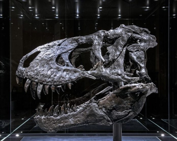
The “Tristan Otto” Tyrannosaurus rex skull that was analyzed by scientists. Credit: RSNA and Charlie Hamm, M.D.
Researchers in Germany determined bone illness in the fossilized jaw of a Tyrannosaurus rex utilizing a CT-based, nondestructive imaging technique, according to a research study existing today at the yearly conference of the Radiological Society of North America (RSNA). The imaging approach might have considerable applications in paleontology, scientists stated, as an alternative to fossil evaluation techniques that include the damage of samples.
A familiar topic these days’s pop culture, the T. rex was an enormous, meat-eating dinosaur that wandered what is now the western United States countless years back. In 2010, a business paleontologist operating in Carter County, Montana, found among the most total T. rex skeletons ever discovered. The fossilized skeleton go back around 68 million years to the Late Cretaceous duration. It was offered to a financial investment lender, who called it “Tristan Otto” prior to lending it out to the Museum für Naturkunde Berlin inGermany It is among just 2 initial T. rex skeletons in Europe.
Charlie Hamm, M.D., a radiologist at Charit é University Hospital in Berlin, and his associates just recently had a chance to examine a part of the Tristan Otto’s lower left jaw. While previous fossil research studies have actually mainly counted on intrusive tasting and analysis,Dr Hamm and associates utilized a noninvasive technique with a medical CT scanner and a strategy called dual-energy calculated tomography (DECT). DECT releases X-rays at 2 various energy levels to offer details about tissue structure and illness procedures not possible with single-energy CT.

CT restorations of the tooth-bearing part of the left dentary. (A) Reconstruction of the standard CT images in lateral view revealing unspoiled physiological structures such as the replacement teeth. The arrow shows the focal exophytic mass– the unusual development that stands out from the surface area of the tissue– on the forward surface area at the level of the 3rd to fifth tooth roots. (B) The DECT-based calcium product map reveals an uniform mineral circulation, while (C) the fluorine product map reveals considerable mineral build-up in the center of the exophytic mass and nearby tooth roots (arrowhead). Credit: RSNA and Charlie Hamm, M.D.
“We hypothesized that DECT could potentially allow for quantitative noninvasive element-based material decomposition and thereby help paleontologists in characterizing unique fossils,”Dr Hamm stated.
The CT method made it possible for the scientists to get rid of the troubles of scanning a big part of Tristan Otto’s lower jaw called the left dentary. The piece’s high density was especially tough, as CT imaging quality is understood to struggle with artifacts, or misstatements of tissue structures, when taking a look at extremely thick things.
“We needed to adjust the CT scanner’s tube current and voltage in order to minimize artifacts and improve image quality,”Dr Hamm stated.
On visual examination and CT imaging, the left dentary revealed thickening and a mass on its surface area that encompassed the root of among the teeth. DECT discovered a considerable build-up of the aspect fluorine in the mass, a finding related to locations of reduced bone density. The mass and fluorine build-up supported the medical diagnosis of tumefactive osteomyelitis, an infection of the bone.
“While this is a proof-of-concept study, noninvasive DECT imaging that provides structural and molecular information on unique fossil objects has the potential to address an unmet need in paleontology, avoiding defragmentation or destruction,”Dr Hamm stated.
“The DECT approach has promise in other paleontological applications, such as age determination and differentiation of actual bone from replicas,” included Oliver Hampe,Ph D., senior researcher and vertebrate paleontologist from the Museum für NaturkundeBerlin “The experimental design, including the use of a clinical CT scanner, will allow for broad applications.”
Dr Hamm and his associates likewise teamed up with paleontologists from the Chicago’s Field Museum and associates from the Richard and Loan Hill Department of Biomedical Engineering at the University of Illinois at Chicago to carry out a CT analysis of the world-famous T. rex “Sue” that is housed in the museum.
“With every project, our collaborative network grew and evolved into a truly multidisciplinary group of experts in geology, mineralogy, paleontology and radiology, emphasizing the potential and relevance of the results to different scientific fields,”Dr Hamm stated.
Additional co-authors are Patrick Asbach, M.D., Torsten Diekhoff, M.D., and Lynn Savic, M.D.
Meeting: 107 th Scientific Assembly and Annual Meeting of the Radiological Society of North America


