Caltech scientists have developed a brand new ultrafast digital camera that may file footage of sign impulses as they journey by means of nerve cells. Credit: Caltech
Reach out proper now and contact something round you. Whether it was the wooden of your desk, a key in your keyboard, or the fur of your canine, you felt it the moment your finger contacted it.
Or did you?
In actuality, takes a little bit of time on your mind to register the feeling out of your fingertip. However, it does nonetheless occur extraordinarily quick, with the contact sign touring by means of your nerves at over 100 miles per hour. In reality, some nerve alerts are even quicker, approaching speeds of 300 miles per hour.
Scientists at Caltech have simply developed a brand new ultrafast digital camera that may file footage of those impulses as they journey by means of nerve cells. Not solely that, however the digital camera can even seize video of different extremely quick phenomena, such because the propagation of electromagnetic pulses in electronics.
Known as differentially enhanced compressed ultrafast images (Diff-CUP), the digital camera know-how was developed within the lab of Lihong Wang. He is the Bren Professor of Medical Engineering and Electrical Engineering, Andrew and Peggy Cherng Medical Engineering Leadership Chair, and government officer for medical engineering.

Lihong Wang. Credit: Caltech
Diff-CUP operates in the same method to Wang’s different CUP programs, which have been proven able to capturing pictures of laser pulses as they journey on the pace of sunshine and recording video at 70 trillion frames per second.
Starting with the identical high-speed digital camera know-how discovered within the different CUP programs, Diff-CUP combines it with a tool referred to as a Mach–Zehnder interferometer. The interferometer pictures objects and supplies by first splitting a beam of laser gentle in two, passing solely one of many cut up beams by means of an object, after which recombining the beams. Since gentle waves are affected by the objects they move by means of, with totally different supplies affecting them in various methods, the beam passing by means of the fabric being imaged may have its waves set out of sync with the waves of the opposite beam. When the beams are recombined, the out-of-sync waves intrude with one another (therefore “interferometer”) in patterns that reveal details about the article being imaged.

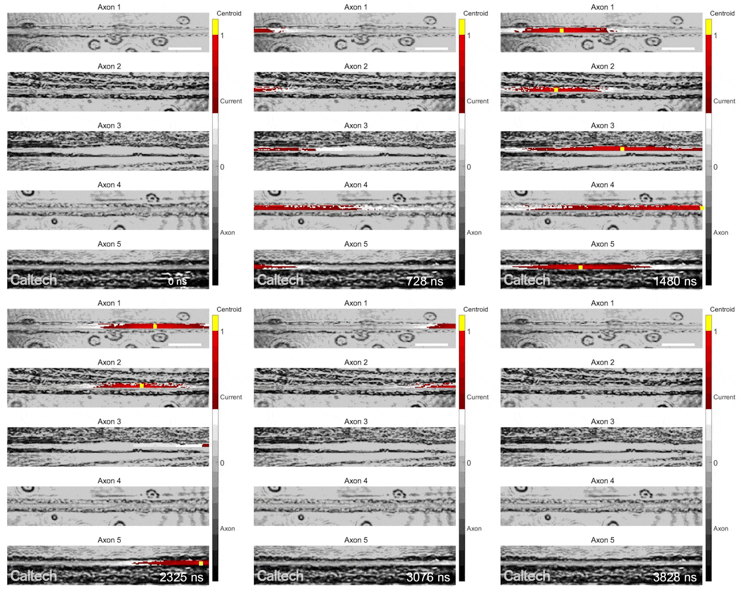
Electrical pulses could be seen touring at totally different speeds by means of totally different neurons. Credit: Caltech
Even although you can’t see {an electrical} pulse touring by means of a nerve cell with your personal eyes, or perhaps a typical gentle microscope, the sort of interferometry can detect it. (Incidentally, this similar primary approach is utilized by LIGO to detect gravitational waves.) So, the Mach–Zehnder interferometer permits the imaging of those pulses, and the CUP digital camera captures the pictures at extremely excessive body charges.
“Seeing nerve signals is fundamental to our scientific understanding but has not yet been achieved owing to the lack of speed and sensitivity provided by existing imaging methods,” Wang says.
Wang’s analysis group additionally captured images of the propagation of electromagnetic pulses (EMP). In some supplies, these can journey at almost the pace of sunshine. In this case, they handed the electromagnetic pulses by means of a crystal of lithium niobate, a salt that has distinctive optical and electrical properties. Despite the extraordinarily excessive pace at which an EMP passes by means of this materials, the digital camera was in a position to clearly picture it.
“Imaging propagating signals in peripheral nerves is the first step,” Wang says. “It’d be important to image live traffic in a central nervous system, which would shed light on how the brain works.”
Reference: “Ultrafast and hypersensitive phase imaging of propagating internodal current flows in myelinated axons and electromagnetic pulses in dielectrics” by Yide Zhang, Binglin Shen, Tong Wu, Jerry Zhao, Joseph C. Jing, Peng Wang, Kanomi Sasaki-Capela, William G. Dunphy, David Garrett, Konstantin Maslov, Weiwei Wang and Lihong V. Wang, 6 September 2022, Nature Communications.
DOI: 10.1038/s41467-022-33002-8
The paper describing their findings appeared in the journal Nature Communications on September 6. Co-authors are Yide Zhang, postdoctoral scholar research associate in medical engineering; Binglin Shen, visitor from Shenzhen University; Tong Wu, visitor from Nanjing University of Aeronautics and Astronautics; Jerry Zhao, former graduate student of the USC–Caltech MD-PhD program; Joseph C. Jing, formerly of Caltech and currently at Cepton; Peng Wang, senior postdoctoral scholar research associate in medical engineering; Kanomi Sasaki-Capela, former research technician at Caltech; William G. Dunphy, Grace C. Steele Professor of Biology; David Garrett, graduate student in medical engineering; Konstantin Maslov, former staff scientist at Caltech; and Weiwei Wang of University of Texas Southwestern Medical Center.
Funding for the research was provided by the National Institutes of Health.


