Scientists have actually made considerable advancements in comprehending brain function utilizing the nematode C. elegans. The research study, using optogenetics and connectomics, uses brand-new insights into neural interaction and difficulties standard designs, improving our understanding of complicated neural networks.
A brand-new research study examines the transfer of details in between nerve cells.
Do we actually understand how the brain works?
In current years, considerable developments have actually been made in understanding the elaborate operations of the brain. Researchers have actually gotten comprehensive understanding about the cellular neurobiology of the brain and have actually discovered a lot about its neural networks and the aspects making up these connections. Despite this, an entire host of crucial concerns stay unanswered and, subsequently, the brain continues to be among science’s terrific, alluring secrets.
Perhaps among the most unpleasant of these concerns focuses on our understanding of the brain as a system. Scientists are still mostly in the dark about how the brain works as a network of engaging elements, about how all the neural elements comply, and specifically, how details is processed in between and amongst this complicated network of nerve cells.
Revolutionary Research on a Simple Organism: The C. elegans Worm
Now, nevertheless, a group of neuroscientists and physicists at < period class ="glossaryLink" aria-describedby ="tt" data-cmtooltip ="<div class=glossaryItemTitle>Princeton University</div><div class=glossaryItemBody>Founded in 1746, Princeton University is a private Ivy League research university in Princeton, New Jersey and the fourth-oldest institution of higher education in the United States. It provides undergraduate and graduate instruction in the humanities, social sciences, natural sciences, and engineering.</div>" data-gt-translate-attributes="[{"attribute":"data-cmtooltip", "format":"html"}]" tabindex =(******************************************************************** )function ="link" >PrincetonUniversity are assisting to shine a clarifying light on how details streams in the brain by studying, of all things, the brain of an extremely little however common worm referred to asCaenorhabditis elegansThe information of the experiment are narrated in a current concern ofNatureThe group includedFrancescoRandi,SophieDvali, andAnujSharma and was led byAndrewLeifer, a neuroscientist and physicist.
“Brains are exciting and mysterious,” statedLeifer“Our team is interested in the question of how collections of neurons process information and generate action.”
https://www.youtube.com/watch?v=7Vy6FerCLqg(**** )
(********************* )Video reveals measurements of neural activity in the worm’s head as private nerve cells are optically promoted one at a time. The nerve cell in the crosshairs is promoted when the words“Stimulated” appear.When nerve cells end up being active they appear dark red in this visualization.The video is accelerated 4x.Credit:FrancescoRandi,PrincetonUniversity
Interest in this concern has broad ramifications,Leifer included.Understanding how a network of nerve cells works is a particular example of a more comprehensive class of concerns in biological physics, specifically, how cumulative phenomena emerge from networks of engaging cells and particles. This location of research study has ramifications for numerous subjects appropriate to biological physics along with modern, advanced innovations, such as expert system.
The primary step in responding to the concern of how details is processed through a network of engaging nerve cells needed that Leifer and his group discover an ideal organism that might quickly be controlled in the laboratory. This ended up being C. elegans, an unsegmented, non-parasitic nematode, or roundworm, that has actually been studied by researchers for years and is thought about a “genetically model organism.” Model organisms are frequently utilized in the lab to assist researchers comprehend biological procedures since their anatomy, genes, and habits are well comprehended.
Innovative Techniques in Brain Mapping and Optogenetics
The worm is roughly one millimeter in length and is discovered in numerous bacteria-rich environments. Especially important to the existing research study is the reality that the organism has a nerve system of just 302 nerve cells in its whole body, 188 of which live in its brain.
“By contrast, a human brain has hundreds of billions of neurons,” statedLeifer “So, these worms are much simpler to study. In fact, these worms are excellent for experimentation because they strike just the right balance between simplicity and complexity.”
Importantly, included Leifer, C. elegans was the very first organism to have its brain electrical wiring totally “mapped.” This indicates that researchers have actually put together an extensive diagram, or “map,” of all its nerve cells and synapses– the locations where nerve cells physically link and interact with other nerve cells. This field of venture is called “connectomics,” in the parlance of neuroscience, and a diagram of an extensive map of neural connections in the brain of an organism is referred to as a “connectome.” One of the primary objectives of connectomics is discovering particular nerve connections accountable for specific habits.

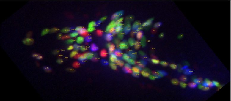
Neurons in the tiny worm C. elegans. The worm’s nerve cells have actually been genetically crafted to be color-coded by their cell type. The names of each nerve cell are displayed in yellow. Credit: Francesco Randi, Princeton University
An extra benefit in utilizing C. elegans in lab experiments is that the worm is transparent, and, in particular cases, its tissue has actually been genetically crafted to be light-sensitive. This location of research study is referred to as “optogenetics” and it has actually reinvented numerous elements of experimentation in biological neuroscience. Instead of the more standard system of utilizing an electrode to provide a present into a nerve cell and therefore promote an action, the optogenetic method includes utilizing light-sensitive proteins from particular organisms and implanting those cells in another organism so that scientists can manage an organism’s habits or reactions utilizing light signals.
Similarly, other proteins can be utilized to illuminate and report when one nerve cell signals to another. This indicates 2 crucial things for lab experimentation: that an organism will react to the existence of light, which a nerve cell, once it gets a signal from another nerve cell, will “light up.” This has actually permitted scientists to study the interaction of nerve cells aesthetically.
“What is really powerful about this tool is that you can literally turn neurons on and watch them signal in real-time,” statedLeifer “In essence, we can convert the problem of measuring and manipulating neural activity to one of collecting and delivering the right light to the right place at the right time.”
These optical tools permitted Leifer’s group to start the painstaking job of comprehending how details streams through the worm’s brain. The objective was to comprehend how signals stream straight through the worm’s whole brain, so each nerve cell needed to be determined. This included separating one nerve cell at a time, shining a light on it, so that it was “activated,” and after that observing how the other nerve cells reacted.
“For this experiment, we went one neuron at a time through the entire brain, activating or perturbing each neuron and then watching the whole network respond,” statedLeifer “This way, we were able to map out how signals flowed through the network.”
“This was an approach that had never been done before at the scale of an entire brain,” included Leifer.
In all, Leifer and his group carried out almost 10,000 stimulus occasions by determining over 23,000 sets of nerve cells and their reactions, a job that took 7 years from conception to conclusion.
Challenging Established Models and Introducing New Insights
The research study carried out by Leifer and his group is so far the most thorough description of how signals stream through the brain. For researchers who study C. elegans, the scientists supplied a great deal of details on how particular signals operate in the worm’s brain, and it is hoped that this research study will offer a huge selection of brand-new details that will assist advance standard research study.
An similarly crucial finding was that a variety of the empirical observations Leifer and his group made throughout the experiment frequently opposed the forecasts of worm habits based upon mathematical designs originated from the worm’s connectome map.
“We concluded that, in many cases, many molecular details that you can’t see from the wiring diagram are actually very important for predicting how the network should respond,” stated Leifer.
The scientists recommend that there is a type of signaling– part of the “molecular details that you can’t see”– that does not advance along neural wires. Leifer and his group identified these as “wireless signals.” Although cordless signaling is popular amongst neuroscientists, it has actually mostly been underappreciated for studying neural characteristics since it had actually frequently believed to be a procedure that happens extremely gradually. Wireless signaling is a type of signaling by which a nerve cell launches particles, called neuropeptides, into the extracellular area, or “extracellular milieu,” in between nerve cells. These chemicals scattered and bind to other nerve cells even if there is no physical connection in between them.
Finally, the scientists think that an essential effect of their work is that it permits other neuroscientists studying this and comparable phenomena to establish much better designs with which to comprehend the brain as a system.
“With our research, we provided a very important piece of the puzzle that was missing,” stated Leifer.
Reference: “Neural signal propagation atlas of Caenorhabditis elegans” by Francesco Randi, Anuj K. Sharma, Sophie Dvali and Andrew M. Leifer, 32 October 2023, Nature
DOI: 10.1038/ s41586 -023-06683 -4
This work was mostly supported by the National Institute of Health New Innovator Award, a National Science Foundation PROFESSION Award, and an award from the SimonsFoundation Funding was likewise gotten from an NSF Physics Frontier Center grant that supports Princeton University’s Center for Physics of Biological Function.


