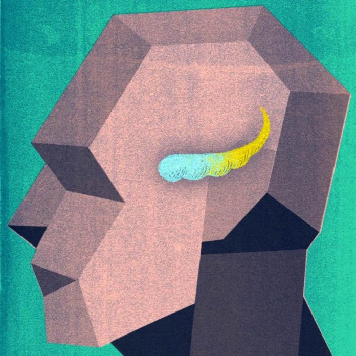
Marked distinctions in gene activity were determined in the anterior part of the hippocampus, which points downward towards the face, and the posterior, which points up towards the back of the head. Credit: Melissa Logies
A research study of gene activity in the brain’s hippocampus, led by UT Southwestern scientists, has actually determined significant distinctions in between the area’s anterior and posterior parts. The findings, released on May 28, 2021, in Neuron, might clarify a range of brain conditions that include the hippocampus and might ultimately assist cause brand-new, targeted treatments.
“These new data reveal molecular-level differences that allow us to view the anterior and posterior hippocampus in a whole new way,” states research study leader Genevieve Konopka, Ph.D., associate teacher of neuroscience at UTSW.

Genevieve Konopka, Ph.D. Credit: UT Southwestern Medical Center
She and research study co-leader Bradley C. Lega, M.D., associate teacher of neurological surgical treatment, neurology, and psychiatry, discuss that the human hippocampus is usually thought about a uniform structure with essential functions in memory, spatial navigation, and guideline of feelings. However, some research study has actually recommended that the 2 ends of the hippocampus – the anterior, which points downward towards the face, and the posterior, which points up towards the back of the head – handle various tasks.
Scientists have actually hypothesized that the anterior hippocampus may be more crucial for feeling and state of mind, while the posterior hippocampus may be more crucial for cognition. However, states Konopka, a Jon Heighten Scholar in Autism Research, scientists had yet to check out whether distinctions in gene activity exist in between these 2 halves.
For the research study, Konopka and Lega, both members of the Peter O’Donnell Jr. Brain Institute, and their associates separated samples of both the anterior and posterior hippocampus from 5 clients who had the structure got rid of to deal with epilepsy. Seizures typically stem from the hippocampus, discusses Lega, who carried out the surgical treatments. Although brain problems set off these seizures, tiny analysis recommended that the tissues utilized in this research study were anatomically regular.

Bradley C. Lega, M.D. Credit: UT Southwestern Medical Center
After elimination, the samples went through single nuclei RNA sequencing (snRNA-seq), which examines gene activity in private cells. Although snRNA-seq revealed mainly the very same kinds of nerve cells and assistance cells live in both areas of the hippocampus, activity of particular genes in excitatory nerve cells – those that promote other nerve cells to fire – differed considerably in between the anterior and the posterior parts of the hippocampus. When the scientists compared this set of genes to a list of genes related to psychiatric and neurological conditions, they discovered considerable matches. Genes related to state of mind conditions, such as significant depressive condition or bipolar illness, tended to be more active in the anterior hippocampus; alternatively, genes related to cognitive conditions, such as autism spectrum condition, tended to be more active in the posterior hippocampus.
Lega keeps in mind that the more scientists have the ability to value these distinctions, the much better they’ll have the ability to comprehend conditions in which the hippocampus is included.
“The idea that the anterior and posterior hippocampus represent two distinct functional structures is not completely new, but it’s been underappreciated in clinical medicine,” he states. “When trying to understand disease processes, we have to keep that in mind.”
Reference: “Resolving cellular and molecular diversity along the hippocampal anterior-to-posterior axis in humans” by Fatma Ayhan, Ashwinikumar Kulkarni, Stefano Berto, Karthigayini Sivaprakasam, Connor Douglas, Bradley C. Lega and Genevieve Konopka, 28 May 2021, Neuron.
DOI: 10.1016/j.neuron.2021.05.003
Other UTSW scientists who added to this research study consist of Fatma Ayhan, Ashwinikumar Kulkarni, Stefano Berto, Karthigayini Sivaprakasam, and Connor Douglas.
This work was moneyed by grants from the National Institutes of Health (NIH grants NS106447, T32DA007290, T32HL139438, NS107357), a University of Texas BRAIN Initiative seed grant (366582), the Chilton Foundation, the National Center for Advancing Translational Sciences of the NIH (under Center for Translational Medicine award UL1TR001105), the Chan Zuckerberg Initiative (an encouraged fund of the Silicon Valley Community Foundation, HCA-A-1704-01747), and the James S. McDonnell Foundation 21st Century Science Initiative in Understanding Human Cognition (scholar award 220020467).


