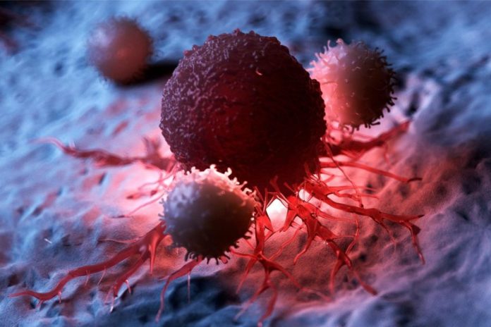
The examine found connections between the PI3K/Akt and p53 pathways that present potential targets for novel most cancers therapies.
An sudden relationship between two of essentially the most frequent cancer-causing components may result in simpler medicine.
According to a latest examine from the University of Wisconsin-Madison, two of the commonest genetic modifications that end in cancerous cells, which have been beforehand believed to be distinct and managed by totally different mobile alerts, are actually working collectively.
To deal with most cancers, researchers have to date targeting growing medicines that both inhibit one or the opposite. Treatments that work higher might consequence from an understanding of their cooperative results.
Cells manufacture a protein referred to as p53, which features throughout the cell nucleus to react to emphasize, however mutations within the gene that makes p53 are the commonest genetic abnormalities in most cancers. Runaway cell proliferation in most cancers can be typically linked to mutations that activate a cell’s surface-located pathway referred to as PI3K/Akt.
Cellular signaling pathways permit cells to perform vital communications duties that keep wholesome cell features. The course of is a bit like sending mail, which requires a selected sequence of steps and applicable stamps and marks on the envelope to ship a letter to the proper deal with.

Outlined in inexperienced, this nucleus of a cancerous cell accommodates DNA in blue and purple blobs marking the cell’s p53 protein binding with components of the Atk mobile signaling pathway, a partnership that can stop the cancerous cell from dying because it ought to and as an alternative extend its life and lead it to divide into extra most cancers cells. Credit: Mo Chen
A crew led by UW–Madison most cancers researchers Richard A. Anderson and Vincent Cryns has found a direct hyperlink between the p53 and PI3K/Akt pathways. The findings, not too long ago printed within the journal Nature Cell Biology, recognized hyperlinks within the pathways that make promising targets for brand new most cancers therapies.
“We have known for some time that lipid messenger molecules that activate the PI3K/Akt pathway found in membranes are also present in the nucleus of cells,” says Anderson, a professor on the UW School of Medicine and Public Health. “But what they were doing in the nucleus separate from membranes was a mystery.”
Mo Chen, an affiliate scientist and first writer of the brand new examine, used chemotherapy medicine to emphasize most cancers cells and injury their DNA as they were replicating or creating new copies of themselves (which cancer cells do often). She discovered that proteins called enzymes that are part of the PI3K/Akt pathway bind to the mutated p53 protein in the nucleus of the cell and attach lipid messengers to p53, showing the two are directly linked.
Instead of entering apoptosis — the proactive process of cell suicide which removes damaged cells — the cancer cells repaired their chemotherapy-damaged DNA and went on growing and dividing, promoting cancer growth.

From left, Vincent Cryns, Mo Chen, and Richard A. Anderson. Credit: Richard A. Anderson, Tianmu Wen
“Our finding that the PI3K/Akt pathway is anchored on p53 in the nucleus was entirely unexpected,” says Cryns, a physician-scientist and professor at UW School of Medicine and Public Health.
The PI3K/Akt pathway was thought to be confined to membranes.
“These results also have critical implications for cancer treatment,” Cryns says. Current treatments that target PI3K may not work because they operate on a different enzyme than the one in the pathway the research team discovered.
The enzyme in the new pathway is called IPMK and rendering it inactive keeps p53 proteins from binding with and activating the Atk pathway, like correcting the address on an envelope so it doesn’t go to the wrong place. This prevents the pathway from benefitting cancer cells, making IPMK a promising new drug target.
The researchers, whose work is supported by the National Institutes of Health, the Department of Defense, and the Breast Cancer Research Foundation, have also identified another enzyme, called PIPKIa, that is a key regulator of both p53 and Akt activation in the cell nucleus.
The team had previously shown that PIPKIa stabilizes the p53 protein, allowing it to be active. When PIPKIa was turned off, p53 levels inside the cell fell sharply. In the new study, the team showed that blocking PIPKIa by genetic approaches or a drug triggered cancer cell death by preventing p53 from activating Akt in the cell nucleus.
“What this means is that drug inhibitors of PIPKIa will reduce mutant p53 levels and block Akt activation in the nucleus, potentially a very powerful one-two punch against cancer cells,” Cryns says. Their team is actively searching for better PIPKIa drug inhibitors that could be used to treat cancers with p53 mutations or abnormally active PI3K/Akt pathway.
In addition to searching for drugs to block the newly discovered cancer pathway, the scientists are investigating whether other proteins in the cell nucleus are targets of the PI3K/Akt pathway.
“We know other nuclear proteins are modified by lipid messengers like p53, but we have no idea how broad the landscape is,” Anderson says.
However, the evidence suggests that this could be a feature shared among many kinds of cancers, “a mechanism we are calling a third messenger pathway,” he adds.
Reference: “A p53–phosphoinositide signalosome regulates nuclear AKT activation” by Mo Chen, Suyong Choi, Tianmu Wen, Changliang Chen, Narendra Thapa, Jeong Hyo Lee, Vincent L. Cryns, and Richard A. Anderson, 7 July 2022, Nature Cell Biology.
DOI: 10.1038/s41556-022-00949-1
This study was funded in part by the National Institutes of Health, the Department of Defense, and the Breast Cancer Research Foundation.


